
Page 9 - KNEETA
P. 9
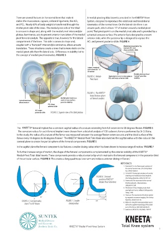
There are several features in the normal knee that make it    A medial-pivoting tibial insert is provided in the KNEETA® Knee
            stable: the musculature, capsule, collateral ligaments, the ACL,    System, designed to reproduce the rotational and translational
            and PCL.  Nearly 60% of body weight is transferred through the    kinematics of the normal knee. On the lateral side there is an
            medial pivot side of the knee.  The medial pivot side of the tibial  arcuate path, which allows 15° of motion around a medial-pivot
            is concave in shape and, along with the medial pivot intercondylar   point. That pivot point is on the medial pivot side and is provided by a
            plateau beminence, acts to prevent anterior translation of the medial  spherical concave surface. The anterior lip is designed to prevent
            pivot femoral condyle.  The opposite is true, however, for the lateral   anterior slide, while the posterior lip is designed to replace the
            compartment of the knee.  This side is convex in shape and,   ACL and prevent posterior slide. FIGURE 4
            coupled with a “humped” intercondylar eminence, allows arcuate              Medial pivot posterior lip
            translation.  These structures create a knee that is more stable on the     replaces ACL and
                  pivot                                                                 stops posterior slide
            the concept of medial-pivot kinematics. FIGURE 3


                                                               Lateral meniscal
                                                               path allows for 15°
                                    A                          of motion
                                                                                                Medial pivot meniscal
                                                               FIGURE 4  |  Medial-  Medial pivot anterior lip replaces   “socket” provides
                                                                 Pivot Insert  PCL and stops anterior slide  stability

                   B         C                 D

                                                               FIGURE 5  |  The KNEETA®
                                                                 femur features spheri-
                                            Medial pivot meniscus   cal femoral condyles
               Lateral meniscus             and concave surface
               allows motion                provide stability
                                   PCL stops
                                   anterior slide
                          ACL stops
                          posterior slide  FIGURE 3 |  Superior view of the tibial plateau


                 KNEETA                                                                               FIGURE 6
               The curvature values for each femoral implant were chosen from a detailed analysis of 130 cadaveric femora performed by Dr D. Blaha.

                                             6   The KNEETA® Medial-Pivot Total Knee also matches the sagittal radius with the radius in the
             coronal plane to create the partial sphere of the femoral components. FIGURE 5
                                                                                                      7
             In the sagittal plane the femoral component also features a smaller closing radius which has been shown to increase range of motion.  FIGURE 7
             To further increase range of motion, the shape of the femoral component is complemented by the anterior stability of the KNEETA®

             Medial-Pivot Total tibial inserts. These components provide a robust anterior lip which maintains the femoral component in the posterior third
             of the articular surface. FIGURE 8                                              1
                                                                                        REFERENCES
                                                                                         1.
                                                                                          the normal human knee. Clin Orthop Relat
                                                                                          Res. 2003;410:69-81.
                                                                                         2.  Schmidt R. Fluoroscopic analyses of cruciate
                                                                     FIGURE 8  |  Femoral   retaining and medial pivot knee implants.
                                                                                          Clin Orthop Relat Res. 2003;410:139-147.
                                                   Closing Radius      position of KNEETA®      3.  Freeman M. The movement of the normal
                                                                       Medial-Pivot Total Knee
                                  90˚                                                     tibiofemoral joint. J Biomechanics.
                                                                                          2005;38:197-208.
                                                                                         4.  Pinskerova V. Knee imaging study sheds
                                 60˚                                                      Today. 1999.
                                                                                         5.  Freeman. The movement of the knee studied
                             30˚                                                          by magnetic resonance imaging. Clin Orthop
                         0˚                                             1/3 A-P
                FIGURE 6  |  Constant radius    FIGURE 7  |  Smaller    A-P               Relat Res. 2003;410:35-43.
                                                  closing radius                         6.  Blaha JD. Using the transepicondylar axis to
                                                                                          part of the femur. JBJS 2002; 84:S48-55.
                                                                                         7.  Iwaki et al, JBJS 82-B, n°8 (2000): 1189-95
                            SUNTEK                                                        KNEETA           R
                            Medical Devices
                            and Electronic
                            Products Trade Co.    KNEETA® Medial-Pivot Knee System     Total Knee system
                                                                                                                 9