
Page 18 - KNEETA
P. 18
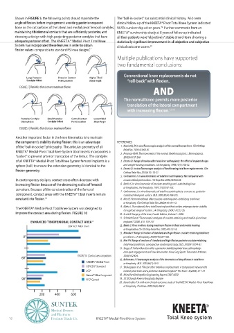
Shown in FIGURE 5, the following points should maximize the   The “ball-in-socket” has substantial clinical history.  Mid-term
                                                            clinical follow-up of the KNEETA® Pivot Total Knee System indicated
         bone on the cut surfaces of the lateral and medial pivot femoral condyles,  96.9% survivorship at ten years.   Further comments from an
                                                                                   19
                                                            KNEETA® survivorship study at 8 years of follow up indicated
         choosing a design with high-projecting posterior condyles that have   all their patients were “objectively” stable at mid-term showing a
                              KNEETA       Pivot Total
                                                            clinical outcome scores.
                                                                             20
                  comparable to standard PS knee designs. 17
                                                             Multiple publications have supported
                                                             two fundamental conclusions:


                                                                Conventional knee replacements do not
               Large Posterior  Posterior Contact  Higher Tibial
                               Point Location  Slope Angle
                                                                               AND
           FIGURE 5
                                                                The normal knee permits more posterior
                                                                  translation of the lateral compartment
                                                                                       1,5-8

            Posterior Condylar  Small Posterior  Central Contact  Lower Tibial
            Osteophytes              Point Location  Slope Angle
           FIGURE 6 |

         Another important factor in the knee kinematics is to maintain
                                                            REFERENCES
         of the “ball-in-socket” philosophy.  The articular geometry of all      1.
                                                                Relat Res. 2003;410:69-81.
         KNEETA® Medial-Pivot Total Knee System tibial inserts incorporates a      2.  Freeman MAR. The movement of the normal tibiofemoral joint. J Biomechanics.
         “socket” to prevent anterior translation of the femur.  The condyles   2005;38:197-208.
         of all KNEETA® Medial-Pivot Total Knee System femoral implants is a      3.
         sphere (ball) to ensure the extension geometry is identical to the   and weight-bearing conditions. J Arthroplasty. 1998;13(7):748-52.
                                                               4.
                                                                Orthop Relat Res. 2003;410:114-21.
                                                               5.
         In contemporary designs, contact areas often decrease with   concave tibial joint surface. J Orthop Res. 2000;18:856-64.
                                                               6.  Banks S. In vivo kinematics of cruciate-retaining and –substituting knee
                                                                arthroplasties. J Arthroplasty. 1997;12(3):297-303.
         curvature. Because of the constant radius of the femoral      7.  Uvehammer J. In vivo kinematics of total knee arthroplasty: concave vs. posterior-
         component, contact areas with the KNEETA® tibial inserts remain   stabilized tibial joint surface. JBJS. 2000;82(4):499-505.
                        18                                     8.  Most E. Femoral rollback after cruciate-retaining and –stabilizing total knee
                                                                arthroplasty. Clin Orthop Relat Res. 2003;410:101-13.
                                                               9.  Blaha J. The rationale for a total knee implant that confers anteroposterior stability
         The KNEETA® Medial-Pivot Total Knee System was designed to   throughout range of motion. J Arthroplasty. 2004;1 (4):22-26.
                                      FIGURE 10               10.  Scott W. Surgery of the knee, Fourth Edition, Volume 1. 2006.
                                                              11.  Schmidt R et al “Fluoroscopic analyses of cruciate-retaining and medial-pivot knee
                 ENHANCED TIBIOFEMORAL CONTACT AREA 18          implants” COOR, 410: 139-147
                                   CONTACT AREA (mm ) 2       12.
                                                                arthroplasties. Clin Orthop Relat Res. 2003;410:131-8.
                                                              13.
             0°                                                 prostheses. J Arthroplasty. 2009;24(5):674-80.
                                                              14.
                                                                total knee prostheses: a prospective randomized study. JBJS. 2009;91:1874-81.
                                                              15.  Suggs JF. Patient function after a posterior stabilizing total knee arthroplasty:
                                                                cam-post engagement and knee kinematics. Knee Surg Sports Traumatol Arthrosc.
            60°                         FIGURE 10 | Contact area comparison    16.   2008;16:290-6.
                                             KNEETA® Medial-Pivot  arthroplasty. JBJS. 2002;84(1):50-3.
                                             GENESIS® Standard    17.  Shakespeare et al “Flexion after total knee replacement. A comparison between the
                                             LCS®               medial-pivot knee and a posterior stabilised implant” The Knee 13 (2006): 371-73
                                             Natural® Ultra Congruent    18.  MicroPort Orthopedics Engineering Report, ER97-0059
            90°                              PFC® Curved      19.  2010 Danish Knee Arthroplasty Register
                                                              20.  Karachalios T. A mid-term clinical outcome study of the KNEETA® Medial- Pivot Total Knee
                                                                arthroplasty. The Knee. 2009;16(6):484-8
                     200              400               600

                           SUNTEK                                                        KNEETA           R

                           Medical Devices
                           and Electronic
      18                   Products Trade Co.     KNEETA® Medial-Pivot Knee System    Total Knee system